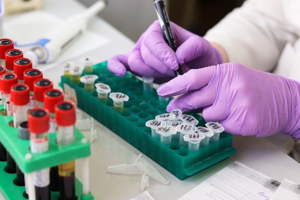
Scientific research is a vast field with various experimentation opportunities carried through the help of different tools to find answers to critical questions. Since a researcher always has endless requirements, keeping track of emerging tools resulting from advanced technology can take time. Every research work can be unique, but everyone needs research instruments. When discussing instrumentation, you refer to intellectual property, software, equipment, and tools. When working with flow cytometry, you prepare a sample for examination. All of them combined help solve mysteries. If you know what research instruments are accessible, you can make concrete decisions and record their usage to help your fellow scientists do their work.
In the context of research instrumentation, one cannot ignore the growing popularity of flow cytometry technology. The laser-based technique helps study particles or cells minutely. You can learn about their chemical and physical compositions in a fluid mixture. Because of its swift analysis of cells’ attributes that enable you to arrive at quantitative and qualitative data, this technology has become an integral part of the research and clinical environment in no time.
Flow cytometry purpose
You can also rely on this technology to examine other suspended particles like molecules and chromosomes in a liquid mixture. Its usage in cell sorting, microorganism detection, health disorder diagnosis (blood cancer, for example), genome size measurement, biomarker detection, and protein development is common. With the help of flow cytometry analyzers, you can gather quantifiable sample data. Cell sorters usually help separate and purify chosen cells per their optical characteristics. The tool allows you to examine particles for fluorescence and visible light scatter during the analysis process. Visible light scatter can be of two types: forward scatter (FSC) and side scatter (SSC). FSC examines the cell’s relative size, while SSC digs into the cell’s granularity. As for fluorescence, it investigates the targeted attributes of the particles labeled as fluorescent antibodies or fluorescent proteins. To be precise, flow cytometry helps with:
- Determining cell size, along with exploring their composition and potential problems
- Evaluating cell’s internal complexity to understand their active state and difference
- Detecting ploidy variations, DNA abnormalities, and others through the measurement of the cell’s DNA
- Assessing DNA synthesis to get an insight into the dynamics of the cell cycle and proliferation
- Quantifying expression levels of the gene in every cell to create distinct cell groups per their molecular profiles
- Detecting and quantifying cell’s surface receptors to understand their functionalities and interactions
- Examining intracellular proteins to get knowledge about cellular responses, protein expression, and more
The working of flow cytometry
As hinted, this technology works on fluorescence and light scattering models. A laser beam passes through the cells or particles in a complex fluid solution to measure the light those particles produce and the fluorescence created during their interactions with certain markers. Light scattering involving FSC and SSC gives insights into cells’ physical characteristics. FSC reveals the surface area of the particles by detecting the spread of light on the forward side. Side-scattered light informs you about the cell’s internal complexity by tracking the light reflection caused by a modified refractive index. SSC and FSC measurements are great for working with heterogeneous cell types.
Fluorescence unravels the cell’s markers or specific molecules. Fluorescent markers or fluorochromes identify nucleic acids or proteins. When laser light passes through the fluorescent compound, it stimulates the electron, which emits fluorescence while returning to its ground state. The tool picks the resulting fluorescence from the fluorescent compound. With the help of the fluorochromes, it distinguishes the cell groups per their fluorescence patterns. You suspend cells retrieved from blood, disaggregated tissues, or cell culture in different tubes for staining while retaining a small number of unstained cells for control.
Flow cytometry components
Optics systems, fluidics systems, and electronics systems are the parts of flow cytometry. The optics system contains collection optics and excitation optics. Excitation optics controls the laser beam’s interaction with the sample, while the other gathers light from the cell-laser interaction. Photodiode and photomultiplier tubes (PMTs), types of optical detectors, catch light signals. Each sensor has a unique wavelength, and filters are there to pick them. The Fluidics system sends the cells through the fluid stream to the laser beam. The flow chamber brings the sample core into the spotlight in the sheath fluid.
However, The electronic system translates the detector’s signals into digital signals for examination. The photomultiplier tubes in the design help convert light signals into electrical waves to amplify and change them into voltage pulses. Then, the Analog-to-Digital Converter translates them into digital numbers for the computer to process and analyze them.
The evolution of flow cytometry instrumentation
When working with flow cytometry, scientists use fluorescence-activated cell sorter (FACS) to sort cells by their unique properties. They choose and gather them in collection vessels for purification. They do sorting through spectral-based techniques, which enable them to segregate particles on their positive and negative attributes. Techniques like high-frequency liquid stream oscillation and charge-based sorting come in handy. To maintain the cell’s integrity, the scientists follow gentle procedures in a sterile environment during sorting. Spectral flow cytometry covers the entire journey of the fluorophore instead of focusing only on the peak levels. The system employs various detectors and methods to segregate the spectra. There is also something called imagery cytometry. It assesses cell morphology and visualizes protein co-expression, immune cell interactions, cell binding, etc.
Reagents are integral to flow cytometry as they enable you to detect and analyze cells and particles. They come in handy with labeling the targeted particles and creating identifiable signals. Nevertheless, flow cytometry is a powerful technology that helps fields like cell sorting, DNA analysis, marine biology, plant biology, virology, microbiology, etc. Many reputable life sciences products and services companies provide these and other lab technologies to make scientists’ work more comfortable and speedy. If you keep yourself upgraded with the latest offerings in the lab field, your research work can become swift and accurate. When you explore options, analyze them based on your research needs and the possibility of wider application.
You may also like
-
Stop the Specialist Shuffle: How a Single Point of Contact Reduces Medical Errors
-
Stop Ignoring Leg Heaviness: How to Identify and Treat Chronic Venous Insufficiency
-
How St George Adults Stop Chronic Joint Pain Without Surgery or Pills
-
High‑Risk Skin Signs Southern Utah Residents Should Never Ignore
-
Ways St. George Residents Can Maintain a Flawless Manicure Between Appointments